Echocardiography
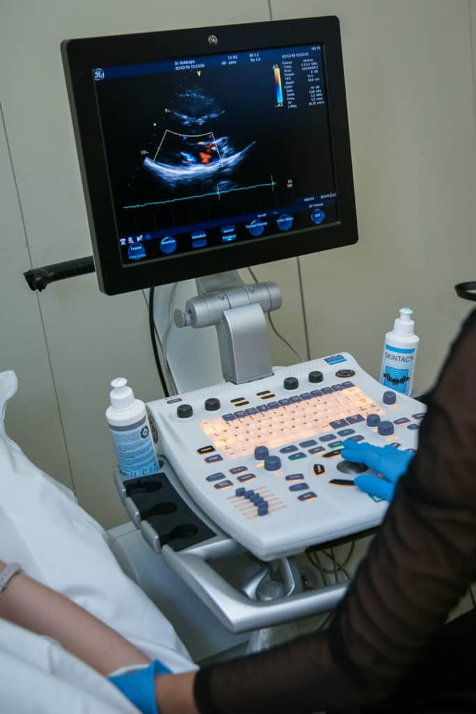
Echocardiography is the study of the heart using ultrasound. Similar to a scan of a baby undertaken during pregnancy, an ultrasound probe applied to the chest wall can be used to study the heart. Ultrasound produces images of the pumping and collecting chambers of the heart, the heart valves, and the main blood vessels, providing detailed information about heart structure and function.
There are several types of scan including:
Transthoracic echocardiography (TTE)
The standard echo (transthoracic echocardiogram) takes about twenty minutes and is performed in the outpatient setting. It is painless and completely safe. A small handheld ultrasound probe is moved over the front of the chest wall to produce images of the beating heart on an adjacent monitor. The size and shape of the various heart chambers can be seen, together with the movement of the heart valves. A vast amount of information can be gleaned from this test, which helps greatly with the diagnosis and management of a wide variety of heart conditions. For example, the effects of a heart attack on the heart muscle can be accurately assessed, the severity of a leaky or narrowed valve can be quantified, and many different causes of breathlessness can be identified.
Stress echocardiography
This is a dynamic form of echocardiography, whereby repeated ultrasound scans of the heart are carried out when the heart is placed under increasing amounts of controlled stress. Exercise in the form of stationary cycling or walking on a treadmill may be used to “stress” the heart and make it beat faster and harder, but in some instances, particularly when a patient is unable to exercise, an infusion of the drug dobutamine is used instead. The purpose of a stress echocardiogram is to highlight where the blood supply to the heart becomes inadequate when placed under stress, which will help with the assessment and management of angina and certain valve problems. In most cases patients are advised to stop drugs called beta blockers five days before the test, since these can prevent the increase in heart rate that typically occurs with stress, but this should only be done on the advice of a cardiologist. The test is an outpatient procedure, taking approximately one hour.
Contrast echocardiography and bubble studies
Contrast echocardiography takes advantage of the acoustic properties of microbubbles in the circulation. Blood conventionally appears black on an ultrasound monitor, because the ultrasound scattered by blood cells at conventional imaging frequencies is very weak. Contrast bubbles oscillating in an ultrasound beam are several million times more effective at scattering sound than red blood cells, and as a result greatly enhance the blood pool signal. This has a number of applications, one of which is to improve the image quality in patients where conventional echo images are suboptimal, for example, due to obesity or lung disease. The contrast is administered as a simple injection into an arm vein.
Contrast can also be used to look for holes and shunts in the heart, such as a patent foramen ovale (PFO) or atrial septal defect (ASD) with an investigation known as a bubble study (shown in the moving image above). As the contrast reaches the right side of the heart, the patient is asked to perform the Valsalva manoeuvre, “bearing down”, which increases the pressure gradient across the heart. If bubbles can be seen flowing from the right atrium into the left atrium (these chambers should be separated completely) then the test result is positive for a hole or shunt. This test takes only slightly longer than a standard transthoracic echocardiogram.
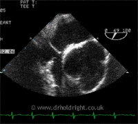
Transoesophageal echocardiography (TOE)
A transoesophageal echocardiogram (TOE) is an ultrasound scan of the heart whereby pictures are taken from inside the body, through the oesophagus rather than from the front of the chest. There are times when the transthoracic echocardiogram cannot provide enough detail, for example in very overweight patients, patients with lung disease and in cases where extremely fine detail is required, particularly when infection of the heart is suspected. The main advantage of a TOE over a transthoracic echocardiogram is that the ultrasound beam does not have to pass through skin, fat, muscle, bone and lung tissue before reaching the heart. The miniaturised TOE probe is attached to a flexible tube which connects to the main echo machine; it is positioned in the oesophagus, which lies behind and immediately adjacent to the heart, giving exquisitely detailed images of the heart structures and function, particularly the areas at the back of the heart, the left atrium and mitral valve, which can be difficult to visualise adequately from the front of the chest.
The TOE is performed in much the same way as an endoscopy. The patient is asked to fast for 6 hours prior to the test, an anaesthetic spray is used to anaesthetise the back of the throat and a sedative is given by injection into an arm vein. The patient lies on their left hand side and the probe is advanced gently into the mouth and then the oesophagus as the patient swallows. It might feel slightly uncomfortable as the probe passes into the oesophagus, but the anaesthetic spray and sedative injection will help to minimise this. The procedure takes about 30 minutes, but because of the sedative the patient will be asked to remain in the department for one to two hours after the test. No fluids or food should be consumed until the local anaesthetic in the throat has worn off, which will again take one to two hours, and due to the sedative driving should be avoided until the following day; it is advisable that a relative or friend drives the patient home after the procedure.
Related links:
Symptoms - Chest Pain
Chest pain has a variety of causes, one of the most important of course being pain from the heart. Read more
Symptoms - Breathlessness and Fluid
Retention
Breathlessness, or dyspnoea, is a common symptom of several medical disorders. Read more
Conditions - Heart Failure
Heart failure develops when the heart can no longer adequately perform its function, which is to pump blood efficiently around the body. Read more
Conditions - Valve Disease
The four chambers of the heart are separated by one-way valves to ensure efficient forward flow of blood from chamber to chamber and into the main blood vessels, the aorta and pulmonary artery. Read more
Conditions - PFO/ASD
The term “hole in the heart” is used to describe a number of conditions, including patent foramen ovale (PFO), atrial septal defect (ASD) and ventricular septal defect (VSD), all of which are present from birth. Read more