Myocardial Perfusion Scanning
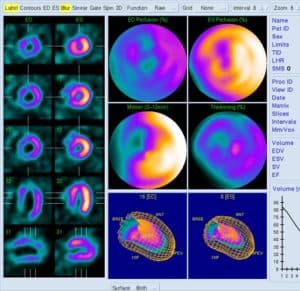
A myocardial perfusion scan is used to evaluate blood flow to the heart muscle and establish how well the heart muscle is functioning. Under stress the heart may not receive enough blood, which can result in symptoms such as angina and breathlessness. As such, in this test the heart is put under a controlled amount of stress, usually in the form of exercise, following which a radioactive tracer called an isotope is injected into a vein, which allows pictures to be produced that show the blood supply to the heart and how well the heart muscle is performing.
The tracer enters heart muscle cells, the amount being proportional to the blood supply the heart muscle is receiving. Areas supplied by a narrowed coronary artery will take up less of the tracer during exercise compared with at rest, giving rise to a “perfusion deficit”, and scarred areas of heart muscle from a previous heart attack will not take up any tracer either at rest or with exercise. As such, the scan gives detailed information about the blood supply to the heart at rest and with stress, and the heart muscle function can also be assessed.
During the test an injection, usually a drug called adenosine, is given into a vein in the arm to dilate the coronary arteries while gentle exercise (pedalling on a stationary bike) is undertaken. Towards the end of the exercise period the tracer, typically technetium or thallium, is injected and approximately 45 minutes later the heart is scanned. During this time the patient will lie still with both arms above the head while the camera moves around the chest, picking up the location and concentration of the isotope in the heart. There is then a break, typically around 90 minutes, following which another radioactive injection may be given. It takes approximately one hour for the tracer to circulate sufficiently to allow the images to be taken and during this time the patient is able to leave the clinic and have a light lunch. Upon return the patient is scanned for a second time. The whole process takes between three and five hours.
In terms of preparation the most important point is to avoid all caffeine-containing drinks and foods for 12 hours prior to the test, as caffeine blocks the actions of adenosine and will therefore reduce the accuracy of the test. It is important to note that caffeine is not only found in coffee and tea, but also chocolate, and some painkillers and cold and ‘flu remedies, amongst other things:
| Food | Caffeine (mg/100ml) | Medicine | Caffeine (mg/pill) |
| Dark Chocolate | 71.4 | Panadol Extra | 65 |
| Milk Chocolate | 21.4 | Solpadeine Plus | 30 |
| Chocolate Cereal | 3 | Benylin Cold and ‘Flu | 25 |
| Drink | Caffeine (mg/100ml) | Drink | Caffeine (mg/100ml) |
| Espresso | 173.6 | Pepsi | 10.7 |
| Fresh Coffee | 45.4 | Green Tea | 10.6 |
| Red Bull | 32 | Diet Pepsi | 10.1 |
| Monster | 32 | Coca Cola | 9.7 |
| Instant Coffee | 24.1 | Chocolate SlimFast | 8.4 |
| Tea | 19.9 | Hot Chocolate | 3.8 |
| Diet Coke | 12.7 | Lipton Iced Tea | 2.8 |
| Lucozade | 12.1 | Decaf Coffee | 2.4 |
| Dr Pepper | 11.8 | Cocoa | 2.1 |
Patients should take along a list of all drugs and inhalers, and dipyridamole (Persantin, Asasantin) should be stopped 48 hours before the scan. After the test, the small amount of radioactive chemicals in the body decay over time and will be excreted in the urine and faeces. Since the scan involves radiation, pregnant women should not have the test, and close contact with pregnant women and children should be avoided after the scan, the time limit depending on the isotope used. Rarely the tracer can be detected by airport security detectors for up to four weeks after the test; patients should ask their doctor to issue them with an explanatory letter if they are planning air travel during this time.
Related links:
Symptoms - Chest Pain
Chest pain has a variety of causes, one of the most important of course being pain from the heart. Read more
Conditions - Coronary artery disease and
angina
Coronary artery disease is the term given to soft fatty deposits or hard calcified plaques within one or more of the coronary arteries, the vessels which supply blood to the heart. Read more
Tests - Coronary Angiogram
Coronary angiography, also called cardiac catheterisation, is a sophisticated test undertaken by an interventional cardiologist as a day case in hospital, which allows the doctor to obtain detailed information about the coronary arteries, and the extent and severity of any disease within them. Read more
Treatments - Coronary Angioplasty/Stent
Coronary angioplasty/stenting, also called percutaneous coronary intervention (PCI) or the balloon and stent procedure, is a treatment used to deal with tight narrowings in the coronary arteries that are affecting the blood supply to the heart. Read more
Treatments - Coronary Artery Bypass
Surgery
Furring up and narrowing of the coronary arteries impairs blood supply to the heart muscle and causes symptoms, typically chest discomfort, which can affect quality of life and, in some cases, prognosis. Read more